Chapter 2 : The Spinal Cord

Brain: Contents Page

The Grey Matter of the Spinal Cord : Ventral Horn

The neurones of the grey matter of the spinal cord are organised into different groupings. The following will be considered in this section:

The Ventral Horn

  • The organisation of motoneurones into longitudinal columns.
The Lateral Horn
  • Autonomic Neurones


Rexed's Laminae : The Arrangement of Neurones within the Spinal Grey Matter

A neuroanatomist called Rexed looked at the structure of the grey matter of the spinal cord and divided it into columns of cells that were similar to each other in different segments.

His observations were based on the shape and size of the neurones, and he divided the grey matter into 10 (Roman X) layers and 6 of these are in the dorsal horn, numbered I to VI .

The classification has been useful in that the different lamina have neurones with different functions.

Neurones involved in Motor or Autonomic Control

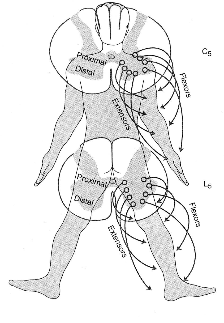
In the ventral horn, lamina VIII and IX contain longitudinal columns of motoneurones. Each column occupies its own position in the ventral horn and extends over several segments of spinal cord.

Extensor motoneurones are arranged in columns nearer the midline of the ventral horn, and flexor columns are situated laterally.


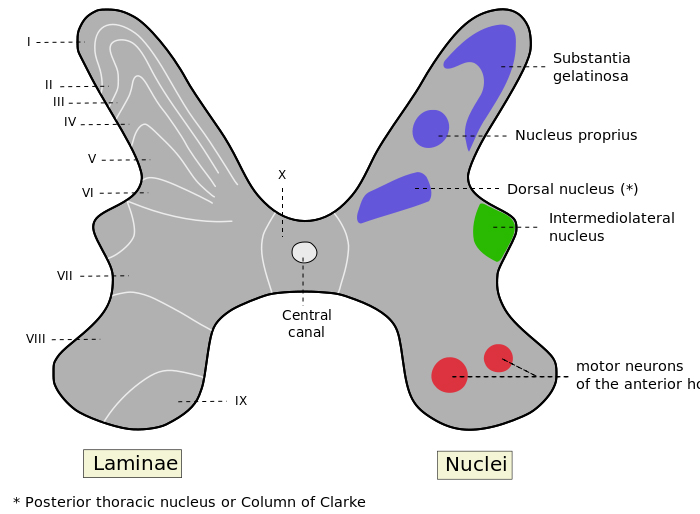
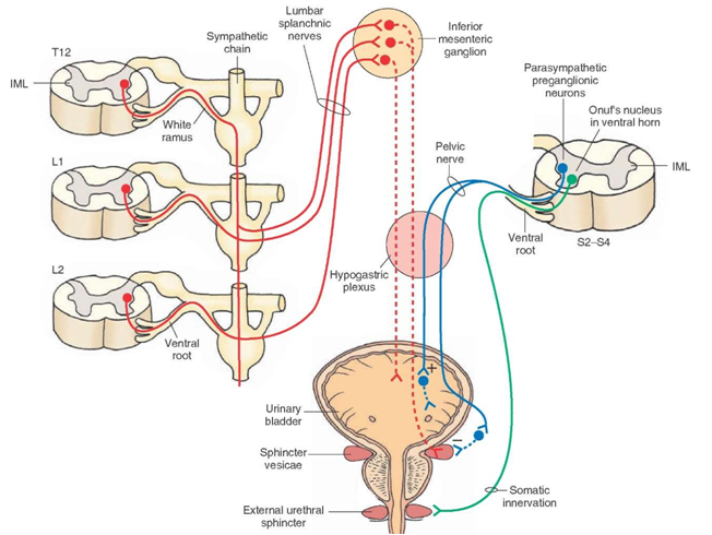
In segments T1 to L2 and the first 4 sacral segments, the grey matter has an intermedio-lateral column of cells which give rise to autonomic pre-ganglionic neurones .

Todd, Nature Neuroscience Reviews
www.neurology.com
http://www.ib.cnea.gov.ar/

Top


Docstoc

The Ventral Horn

Motoneurones and Interneurones

In the ventral horn, Lamina VIII and IX contain motoneurones of different types.

Flexor and Extensor Motoneurones

The diagram shows that the medial column (IX) controls extensors amd the lateral column controls flexors.

Proximal and Distal Muscle Innervation

There is a further topographic arrangement in that within any segment, the most ventral motoneurones innervate distal muscles and the motoneurones that occupy a more dorsal position in the ventral horn innervate proximal muscles in the limb.

Rostral vs Distal Segmental Arrangements

In addition, the proximal muscles of each limb are controlled by the rostral segments and the distal muscles are connected to motoneurones in lower parts of the cord innervating the limb.


Motor neurons that innervate flexor muscles are located dorsal to motor neurons that innervate extensor muscles.

Motor neurons that innervate distal muscles are located lateral to motor neurons that innervate proximal and axial muscles.


Top
The Lateral Horn (the autonomic nucleus in the lateral part of Lamina VII)

Autonomic Pre-ganglionic Neurones

The intermediolateral nucleus (IML) is found in the lateral horn of the spinal grey matter, which is part of Rexed lamina VII. The lateral horn exists only in certain segments of the spinal cord, viz the first thoracic to the second lumbar segments (the sympathetic outflow), and from the second to the fourth sacral segments (the parasympathetic outflow).

Autonomic preganglionic neurones have finely myelinated axons that enter the ventral roots, and proceed towards either the sympathetic or parasympathetic ganglia.

The slide opposite shows a pronounced lateral horn at the level of the central canal.


The diagram opposite shows the difference between the paths of the sympathetic preganglionic neurones, which enter the sympathetic chain, and the parasympathetic neurones that innervate the bladder.

These neurones innervate the lower urinary tract.

In addition the sacral cord has a specialised group of motoneurones that innervate the skeletal muscle of the external sphincters, and the cell bodies of these motoneurones are in a part of the ventral grey matter called "Onuf's nucleus". These neurones have close functional connections with the parasympathetic system, both of which are involved in the control of continence.


Top


Chapter 2 : The Spinal Cord

Brain: Contents Page

© HumanPhysiology.Academy 2014-2015